Welcome to the Tesla Memorial Society of New York Website
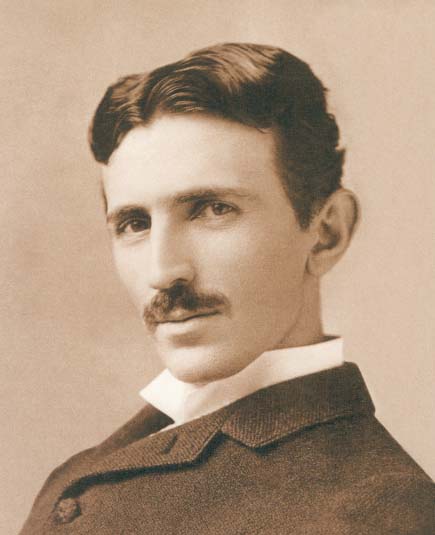
Above: Nikola Tesla (1856-1943) at the age of 38.
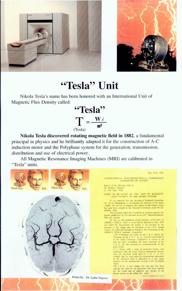
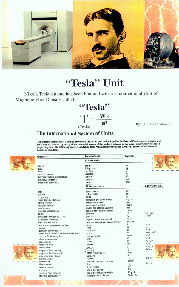
All MRI (Magnetic Resonance Images) machines are calibrated in Tesla Units. Nikola Tesla's name has been honored with the international unit of magnetic flux density called "Tesla"
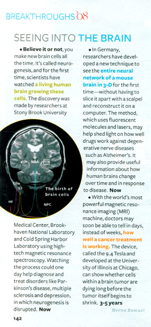
Above: "Seeing into the Brain", Reader's Digest, March 2008.
A Short History of the Magnetic Resonance Imaging (MRI)
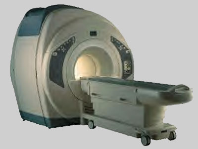
Above: All MRI machines are calibrated in "Tesla" Units.
Nikola Tesla discovered the Rotating Magnetic Field in 1882 in Budapest, Hungary. This was a fundamental discovery in physics.
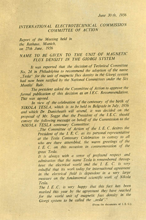
In 1956, the "Tesla Unit" was proclaimed in the Rathaus of Munich, Germany by the International Electro-technical Commission-Committee of Action. All MRI machines are calibrated in "Tesla Units". The strength of a magnetic field is measured in Tesla or Gauss Units. The stronger the magnetic field, the stronger the amount of radio signals which can be elicited from the body's atoms and therefore the higher the quality of MRI images.
1 Tesla = 10,000 Gauss
Low-Field MRI= Under .2 Tesla (2,000 Gauss)
Mid-Field MRI= .2 to 0.6 Tesla (2,000 Gauss to 6,000 Gauss)
High-Field MRI= 1.0 to 1.5 Tesla (10,000 Gauss to 15,000 Gauss)
In 1937, Columbia University Professor Isidor I. Rabi working in the Pupin Physic Laboratory in Columbia University, New York City, observed the quantum phenomenon dubbed nuclear magnetic resonance (NMR). He recognized that the atomic nuclei show their presence by absorbing or emitting radio waves when exposed to a sufficiently strong magnetic field.
Professor Isidor I. Rabi received the Nobel Prize for his work. He is one of 28 Nobel Laureates from the Pupin Physics Laboratory in New York City.
Raymond Damadian, a physician and experimenter working at Brooklyn's Downstate Medical Center discovered that hydrogen signal in cancerous tissue is different from that of healthy tissue because tumors contain more water. More water means more hydrogen atoms. When the MRI machine was switched off, the bath of radio waves from cancerous tissue will linger longer then those from the healthy tissue.
In 1973, Paul Lauterbur, a chemist and an NMR pioneer at the State University of New York, Stony Brook, produced the first NMR image.
Mike Goldsmith, one of the graduate students cobbled a wearable antenna coil to monitor the hydrogen broadcast detected by the coil.
On July 3, 1977, nearly five hours after the start of the first MRI test, the first human scan was made as the first MRI prototype.
How MRI works?
Magnetic Resonance Imaging is a medical diagnostic technique that creates images of the human body using the principle of nuclear magnetic resonance. It can generate thin-section images of any part of the human body - from any angle and direction. MRI is possible to make such a picture of the human body when the body is exposed to an electromagnetic field.
MRI creates a strong magnetic field and the small biological "magnets" in the human body consisting of protons located in the nucleus of the hydrogen atom are magnetized. The proton possess fundamental magnetic properties.
First, MRI creates a steady state of magnetism within the human body by placing the body in a steady magnetic field. Second, the MRI stimulates the body with radio waves to change the steady-state orientation of protons. Third, the MRI machine stops the radio waves and registers the body's electromagnetic transmission. Fourth, the transmitted signal are used to construct internal images of the body by computerized axial tomography.
An MRI image is not a photograph. It is actually a computerized map or image of radio signals emitted by the human body. MRI is superior to CAT scan because CAT scan is using ionizing radiation, MRI uses harmless radio waves. The only unusual preparation is that all removable metallic objects must be left outside the scanning room, including removable hearing aids, dentures and other prosthetic devices. Credit cards can be damaged by the MRI because magnetic codes can be affected by the MRI magnet.
Magnetic Resonance Imaging is a powerful diagnostic tool in the medical imaging market place as the procedure of choice for the visualization of soft tissue. The MRI industry is producing over 2,000 units per year. The United States is represented with 40% of the world marketing and production of MRI. There is an emerging consensus that the MRI has a broad application in smaller hospital and clinics.
Neurologists are one of medical specialty that depend a great deal on MRI for accurate diagnostic of the central nerve system. Other medical specialty that rely upon the MRI technology include neurosurgeons, orthopedic surgeons and chiropractors. MRI is useful in diagnosis of "pinched nerves" in the spinal column, heart disease, multiple sclerosis and other diseases of the central nerve system.
The use of MRI technology will increase in the United States and the world because of the tremendous significance in modern medical diagnosis. The Tesla Unit is a label on every MRI machine signifying the strength of the MRI Magnetic Field. The stronger the magnetic field, the better the image of the MRI. Tesla's name connected with MRI technology will be more and more widely known around the world.
Tesla Posters - Tesla Unit
by Dr. Ljubo Vujovic,
Secretary General, Tesla Memorial Society of New York